What Are the organs in the nervous system, and what do they do?
There are several different organs contained in the nervous system and each is responsible for a different function.
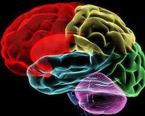
Brain: The brain is the control center of the body. It consists of three main components: the forebrain, the brainstem, and the hindbrain.
The forebrain is responsible for a variety of functions including receiving and processing sensory information, thinking, perceiving, producing and understanding language, and controlling motor function. The forebrain contains structures such as the thalamus and hypothalamus which are responsible for such functions as motor control, relaying sensory information, and controlling autonomic functions. It also contains the largest part of the brain, the cerebrum.
The midbrain and the hindbrain together make up the brainstem. The midbrain is the portion of the brainstem that connects the hindbrain and the forebrain. This region of the brain is involved in auditory and visual responses as well as motor function.
The hindbrain extends from the spinal cord and contains structures such as the pons andcerebellum. These regions assist in maintaining balance and equilibrium, movement coordination, and the conduction of sensory information. The hindbrain also contains the medulla oblongata which is responsible for controlling such autonomic functions as breathing, heart rate, and digestion.
spinal cord
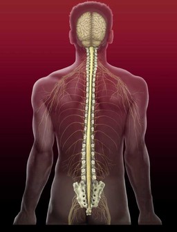
Spinal Cord: The spinal cord is a cylindrical shaped of nerve fibers that is connected to the brain. The spinal cord runs down the centre of the protective spinal column extending from the neck to the lower back. Spinal cord nerves transmit information from body organs and external stimuli to the brain and send information from the brain to other areas of the body. The nerves of the spinal cord are grouped into bundles of nerve fibers that travel in pathways. These fibers carry sensory information from the body to the brain. Descending nerve tracts send information pertaining to motor function from the brain to the rest of the body.
sensory organ
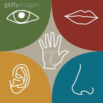
The sense organs — eyes, ears, tongue, skin, and nose — help to protect the body. The human sense organs contain receptors that relay information through sensory neurons to the appropriate places within the nervous system.
Each sense organ contains different receptors.
Eye(s) – for sight:
When you look at an eye, the iris is the colored part. The iris actually is a pigmented muscle that controls the size of the pupil, which dilates to allow more light into the eye or contracts to allow less light into the eye. The iris and pupil are covered by the cornea.
Two types of sensors detect light:
Ear(s) – for hearing and balance:
The ear consists of three main segments:
Olfactory organ – for smell:
This is the organ of smell. It consists of two parts: the external nose, and the internal nasal cavity, which is divided by a septum with right and left nasal chambers. Olfactory cells, which are lined with cilia, line the top of your nasal cavity. Scents (made up of various chemicals) bond to the various cilia which generate a nerve impulse that is carried through the olfactory cell, into the olfactory nerve fiber, up to the olfactory bulb and to your brain. Your brain determines what you are smelling.
Tongue (Taste buds) – for taste:
The senses of smell and taste work closely together. If you cannot smell something, you cannot taste it, either. However, the chemoreceptors in the nose will detect any kind of smell, whereas there are four different types of taste buds, and each detects different types of tastes: sweet, sour, bitter, and salty. The taste buds are located at the base of the tongue and on the floor of the mouth. They contain nerve endings from the glossopharyngeal nerve. Small quantities of the chemicals of taste are recognized by the taste buds and this information is transferred to the appropriate receptors of the brain.
Skin- for touch:
The skin contains general receptors. These receptors can detect touch, pain, pressure, and temperature. Throughout your skin, you have all four of these receptors interspersed. Skin receptors generate an impulse when activated, which is carried to the spinal cord and then to the brain.
Each sense organ contains different receptors.
- General receptors are found throughout the body because they are present in skin, visceral organs (visceral meaning in the abdominal cavity), muscles, and joints.
- Special receptors include chemoreceptors (chemical receptors) found in the mouth and nose, photoreceptors (light receptors) found in the eyes, and mechanoreceptors found in the ears.
Eye(s) – for sight:
When you look at an eye, the iris is the colored part. The iris actually is a pigmented muscle that controls the size of the pupil, which dilates to allow more light into the eye or contracts to allow less light into the eye. The iris and pupil are covered by the cornea.
Two types of sensors detect light:
- Rods detect motion. The rods work harder in low light.
- Cones detect fine detail and color. The cones work best in bright light. There are three types of cones: one that detects blue, one that detects red, and one that detects green. Color blindness occurs when one type of cone is lacking.
Ear(s) – for hearing and balance:
The ear consists of three main segments:
- The outer ear
- The middle ear separated from the outer by the tympanic membrane (ear drum)
- The inner ear consisting of the cochlea and the three semicircular ducts
Olfactory organ – for smell:
This is the organ of smell. It consists of two parts: the external nose, and the internal nasal cavity, which is divided by a septum with right and left nasal chambers. Olfactory cells, which are lined with cilia, line the top of your nasal cavity. Scents (made up of various chemicals) bond to the various cilia which generate a nerve impulse that is carried through the olfactory cell, into the olfactory nerve fiber, up to the olfactory bulb and to your brain. Your brain determines what you are smelling.
Tongue (Taste buds) – for taste:
The senses of smell and taste work closely together. If you cannot smell something, you cannot taste it, either. However, the chemoreceptors in the nose will detect any kind of smell, whereas there are four different types of taste buds, and each detects different types of tastes: sweet, sour, bitter, and salty. The taste buds are located at the base of the tongue and on the floor of the mouth. They contain nerve endings from the glossopharyngeal nerve. Small quantities of the chemicals of taste are recognized by the taste buds and this information is transferred to the appropriate receptors of the brain.
Skin- for touch:
The skin contains general receptors. These receptors can detect touch, pain, pressure, and temperature. Throughout your skin, you have all four of these receptors interspersed. Skin receptors generate an impulse when activated, which is carried to the spinal cord and then to the brain.
